We fix the cornea shape by high frequency irradiation.
The conical cornea latest treatment which it does not cut.
IWe start the advanced treatment mole cricket flextime of the conical cornea.
If it is this latest cure, even the extreme conical cornea state that is diagnosed if there is not a method lets you normalize cornea shape, and improvement of the vision by the naked eye eyesight can expect only corneal transplant.
Please talk about the one of the trouble with a conical cornea.
Keraflex® delivers low energy, high frequency (radio frequency, 915 MHz) energy directly onto the corneal surface through a capacitively coupled Keraflex® applicator. A circular electrode in the applicator delivers energy, in an annular pattern, to the superficial corneal stroma (250 microns below the surface) predictably and homogeneously heating the collagen. This pattern of heating creates a toroidal shrinkage zone with a dose-dependent circular cross-section. This annular treatment of the superficial cornea and the resultant toroidal pattern produces a band of tightening that predictably decreases the curvature of the cornea, over a range of 0.5 to 6.0D. This flattening results in a safe, predictable, and temporary modification to the curvature of the cornea, thereby correcting the point of focus where light impinges onto the retina.
The Vedera KXS is a microwave-based system for reshaping the cornea. Depending upon the energy delivered and the physical dimensions of the microwave emitter, tissue lesions of varying dimensions and positions can be generated through tissue heating. The Vedera KXS performs a circumferential shrinkage using a circular activation of a ring. The amount of shrinkage and change in corneal curvature is proportional to the total applied energy.
The Vedera KXS had received a CE Mark for the following intended use:
DEVICE DESCRIPTION
The Vedera KXS delivers a short burst of energy in a predetermined pattern onto a human cornea. Depending on the energy delivered and the physical dimensions of the antenna, tissue lesions of varying dimensions and positions are generated. The depth of these lesions within the corneal tissue is controlled by actively cooling the surface of the cornea prior to treatment. As a result of the heating, the reorganization of collagen fibers in the stromal tissue results in mechanical stresses, causing a central flattening of the cornea, thus flattening the cornea.
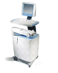
Vedera KXS
The Vedera KXS, shown in Figure 1, consists of the generator console and articulating arm. The Vedera KXS Console houses the system controls, treatment energy generator, power supply, coolant system, vacuum system, built-in printer, and LCD display. It also contains the Articulating Arm, which is an extension of the console; the Articulating Arm provides low-loss, high-reliability transfer of energy and coolant to a location close to the operating field. It also contains the eye ring and targeting brake vacuum lines, DC power and signals, and treatment energy and coolant connectors.
Eye Targeting Stage
The Eye Targeting Stage has two functions 1) immobilize the eye before and during treatment, and provides a rigid mechanical interface to the eye for the targeting stage and the applicator. 2) When reticle is in place and prior to locking the stage position, adjustment of the x-y position of the applicator relative to the eye to align the treatment center with ocular features (pupil center or apex). Figure 3 below illustrates the Eye Targeting Stage.
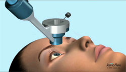
Treatment Applicator
The Treatment Applicator delivers energy and coolant, is part of the mechanical positioning system for the eye (together with the targeting stage), and determines the geometry of the treatment. Its primary function is to deliver a burst of energy to the cornea. The Treatment Applicator includes electrodes for delivery of energy to the cornea, conduits for coolant dispersal above the corneal surface to provide cooling, and feedback controlled motors that position the electrodes for treatment of corneal tissue. To control the z position (depth) of the tissue heating and to cool the cornea’s surface, a controlled amount of liquid evaporative coolant (R-134a) is delivered to the cornea immediately prior to treatment. The Treatment Applicator also contributes to the mechanical control of the eye during treatment by assuming a controlled and locked position relative to the targeting stage.
The Treatment Applicator is held by the ophthalmologist and is placed into the target stage, from where motors advance it onto the eye. A Bioinsulator encloses the electrodes and provides a sterile dielectric interface to the patient’s eye. This applicator cover is single-use.
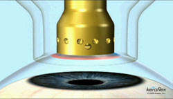
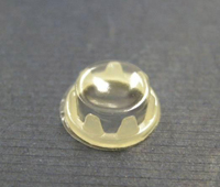
Figure 5 shows the underside of the Treatment Applicator. The outer electrode is the silver ring and the inner electrode is the thin, inner yellow ring. The energy travels between the electrodes, through the cornea of the patient. Note that there is a gap between the electrodes as part of the design.
A typical treatment may be 10-200W of power at a frequency of 915 MHz, with heating pulse duration of 10-100ms. A cooling pulse is typically applied before the heating pulse; further pulses are then provided during the cycle. The purpose is to provide adequate surface cooling of the eye, balanced with the heating pulse to shrink collagen just below the surface of the cornea. The system has several safety features, including sensors that detect coolant flow and pressure, the amount of power, and assure good eye contact.
A Locking Mechanism positively locks the applicator to the targeting stage. The Locking Mechanism engages automatically when the applicator is inserted into the targeting stage.
The applicator has a mechanism to move the treatment tip towards the cornea. The mechanism is enabled only if the applicator is locked to the targeting stage and the footswitch is depressed. The applicator can be removed from the target stage by simultaneously depressing the two black tabs on the sides of the applicator.
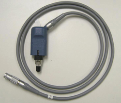
BioInsulator
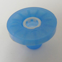
A sterile, single-use BioInsulator (Figure 6) is provided for each treatment to isolate the eye from the applicator electrode, eliminating the risk of cross-contamination between patients. The BioInsulator fits over the treatment applicator; it is applied to the treatment applicator prior to treatment.
The BioInsulator consists of a thin, rigid or flexible dielectric membrane. The BioInsulator prevents direct electrical contact to the eye, allowing for capacitive coupling of the energy. In addition, the membrane forms a protective barrier to prevent the coolant from contacting the eye. The BioInsulator is single-use and a ring can be observed on the cover where the energy was transmitted.
Reticle
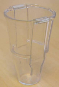
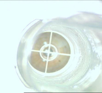
The clear, plastic Reticle (Figure 8) is used to align the targeting stage to the eye. The Reticle contains a cross-hair which is aligned with a mark placed on the corneal surface. This allows for the accurate centration of the treatment applicator over the apex of the cornea.
Exclusion Criteria
All patients meeting any of the following criteria will be excluded from treatment:
1. Eyes classified as normal on the keratoconus severity grading scheme.
2. Corneal pachymetry <400 microns at diameter to be treated.
3. Previous ocular condition (other than refractive error) in the eye(s) to be treated
that may predispose the eye for future complications, for example:
a. History of corneal disease (e.g., herpes simplex, herpes zoster keratitis, recurrent erosion syndrome, corneal melt, corneal dystrophy, etc.)
b. Clinically significant corneal scarring in the treatment zone that is not related to keratoconus or, in the physician’s opinion, will interfere with the treatment.
4. A history of chemical injury or delayed epithelial healing in the eye(s) to be treated.
5. Diagnosed with autoimmune disease, systemic connective tissue diseases or atopic syndrome, diabetes mellitus, or taking systemic medications likely to affect wound healing.
6. Women who are pregnant or nursing
Treatment Preparation
1. Topical anesthetic should be applied per the normal routine of the physician.
2. Keratoconus procedures generally should be centered on the geometric center of the cornea. The physician should mark geometric center of cornea either at the slit lamp or under the operating microscope.
3. The operator should program the system with the subject’s demographic information including name, patient ID, gender, date of birth, and procedure date.
4. The subject’s manifest refraction and keratometry readings should be reviewed and the treatment plan identified.
Treatment
1. During the setup phase, the system will perform internal safety checks.
2. While the patient is in a reclining position, the eye is prepared for treatment according to the physician’s standard procedure. A wire lid speculum of the physician’s choice is placed.
3. The physician will program the system for the desired attempted correction based on the manifest refraction spherical equivalent.
4. The physician will position the targeting stage on the eye to be treated and will enable a vacuum output to hold the targeting stage in place.
5. The physician will insert the cross-hair target reticle into the targeting stage which they manually position over the cornea in alignment with the corneal apex. The targeting stage must be locked in place and the target removed.
6. The physician will insert the applicator into the targeting stage. Once engaged a tone will sound.
7. The physician depresses the footswitch.
8. After the application is complete, the vacuum to the targeting stage is automatically disabled, resulting in detachment of the targeting stage and applicator from the eye.
9. The physician should then remove the targeting stage from the eye.
10. Remove the epithelium across a 9 mm diameter in preparation for cross linking.
11. Proceed with cross linking per the physician’s standard treatment regimen.
The second eye may be treated at the discretion of the physician. Retreatments may be performed after the 1 month examination. Contact lens use should not be resumed for 3 months after the treatment.
Other standard of care therapeutic treatments for keratoconus may be utilized in addition to the Keraflex® treatment.
Keraflex® Animation
Vedera KXS(Figure1)
Eye Targeting Stage(Figure 2)
Microwave(Figure 3)
Treatment Applicator(Figure 4)
Shows the underside of the Treatment Applicator(Figure 5)
BioInsulator(Figure 6)
Cover with Applicator Tool(Figure 7)
Reticle(Figure 8)
Reticle Positioned Over the Eye in Targeting Stage(Figure 9)